
- Hospital Furniture
- Neonatal Care Product
- Operating Surgical Tables
- Orthopedic Implants
- Surgical Operating Led Lights
- Orthopedic External Fixators
- Laryngoscopes
- Orthopaedic Power Tools
- Medical Disposables
- Hospital Clothing
- Anaesthesia Products
- Autoclaves & Sterilizers
- Hospital Holloware
- Suction Units
- Rehabilitation Aids
- Stethoscopes
- Anesthesia Machine & Ventilators
- Cold Chain Equipment
- Electro Medical Equipments
- Orthopedic Instruments
- Surgical Operating Led Lights
- Physiotherapy products
Why Are Dental Equipment Tools Important for Clinics?
A clinic’s care is shaped by more than the clinician’s skill. The instruments on the tray, the suction beside the chair, and the light above the patient all affect how smoothly a procedure can proceed. Dental Equipment Tools help clinicians examine, clean, restore, and monitor oral health. Their importance is practical, not cosmetic.
Dental researcher and prosthodontist Dr. Gordon Christensen has written extensively about evaluating dental products for clinical use. A fair paraphrase of that approach is: “Choose equipment for reliable performance in real clinical conditions, not for appearance alone.” This is a paraphrase, not a verbatim quotation. It points to a useful question for clinic owners: does a tool perform consistently, feel manageable, and fit the team’s workflow? Small details matter. A handpiece that vibrates unexpectedly or a curing light with a weak battery can interrupt treatment and require attention.
Good equipment does not guarantee good outcomes. It must be selected for the procedures a clinic actually provides, maintained according to manufacturer instructions, and used by trained staff. Costs matter, too; the most advanced model may not be the right choice for every practice. That deserves a second look. Clinics can compare durability, service support, ergonomics, and cleaning requirements before purchasing. When these factors are considered together, Dental Equipment Tools can support safer routines, clearer examinations, and more dependable day-to-day care.
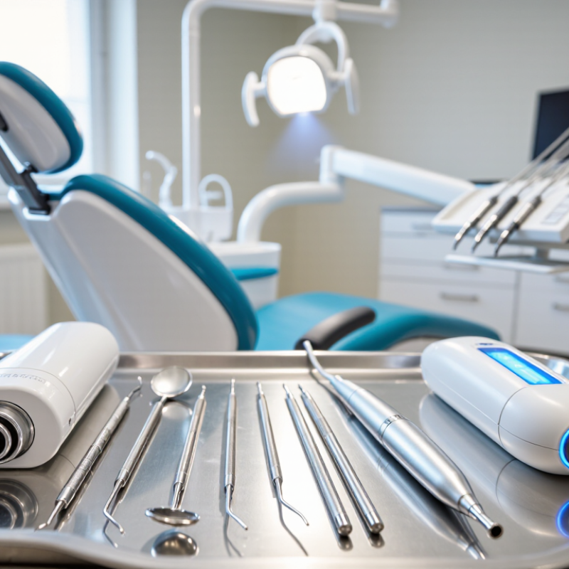
Table of Contents [Hide]
Types of Dental Equipment Used in Clinics
Dental clinics rely on several kinds of equipment, each serving a distinct part of patient care. The dental chair supports positioning, while an adjustable operating light helps clinicians see small details clearly. Handpieces, mirrors, probes, and scalers are used during examinations and procedures. Even a simple mirror matters. It helps reveal areas that are difficult to view directly.
Instruments must be cleaned and sterilized according to established infection-control procedures. A sterilizer, sealed instrument packs, and a clear workflow help staff handle reusable tools safely. Suction equipment removes saliva and water during treatment, keeping the working area more visible. Dental imaging systems can help clinicians assess teeth and surrounding structures when indicated. Their use should follow appropriate clinical protocols, with equipment maintained and images interpreted by qualified professionals.
Clinics also use curing lights for restorative materials and mixing or dispensing tools for selected procedures. The exact setup varies with the services offered, room layout, and patient needs. Equipment choice is not only about having more devices. Staff need training, routine checks, and enough space to move comfortably. A crowded room can slow work, even when every tool is modern. No arrangement is perfect; teams may need to adjust placement after noticing awkward reaches or delayed instrument handoffs.


